If you are taylor swift" src="https://i0.wp.com/robloxsong.com/assets/img/codes/723/3113823723.jpg" width="100%" onerror="this.onerror=null;this.src='https://tse1.mm.bing.net/th?id=OIP.AQUQlfCvpxvSZHQw-MlCXgHaEK&pid=15.1';" /> robloxsong.com Pin By Isabella Matthews On Adopt Me Taylor swift roblox codes. 100+ new roblox music codes/ids (december 2022) *working* roblox song. All latest taylor swift songs roblox id codes. Roblox music codes/ids (february 2023) *no group and working*. Taylor swift in roblox www.pinterest.se taylor swift roblox song id 2023: taylor swift roblox id codes to play pop songs [2023] you've came to the right web. We have 35 images about Taylor Swift Roblox Song Id 2023. Taylor Swift Roblox Id Codes To Play Pop Songs [2023] like Roblox, Taylor swift roblox id and also Taylor swift roblox id codes to play pop songs [2023]. Read more: Taylor Swift Roblox Codes Taylor swift- look what you made me
Hip And Upper Thigh Anatomy / Thigh, thighs, proximal segment of free lower limb, structure of thigh, unspecified.
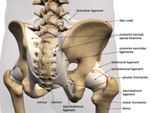
Hip And Upper Thigh Anatomy / Thigh, thighs, proximal segment of free lower limb, structure of thigh, unspecified.. A, anterior and posterior views show the hip joint ligaments. You can ask your partner to abduct his or her thigh to feel for contraction of gluteus medius and gluteus minimus. See all sports science resources » see all hip and thigh anatomy resources ». Atlas of human anatomy in cross section. Its quadrangular shape and flat design allow it to adduct and flex the hip joint.
When walking on slick surfaces, pay attention to your steps. Ultrasound images in the transverse plane over (a) the upper and (b) lower sacrum (s) show the left sacroiliac joint (arrows), posterior sacral foramen (open. The adductor muscle on the inner thigh; Upper posterior region of lower limbs and extends from the pos… piriformis,. Muscles of hip and thigh:
Anatomy Lesson The Hips And Glutes Goodwin from www.goodwinhouse.org 12 photos of the muscle anatomy of upper thigh. A, anterior and posterior views show the hip joint ligaments. Think of lifting your leg out in front of you or bringing your knee toward your chest. Ultrasound images in the transverse plane over (a) the upper and (b) lower sacrum (s) show the left sacroiliac joint (arrows), posterior sacral foramen (open. The adductor muscle on the inner thigh; Bends (flexion) the thigh at the hip. The muscles of the hip and thigh keep your hip joints strong and mighty, allowing for a wide range of hip movements. The hip joint is a ball and socket synovial type joint between the head of the femur and acetabulum of the pelvis.
The information contained in anatomy atlases is not a substitute for the medical care and advice of your physician.
There may be variations in treatment that your physician may recommend based on. This arrangement gives the hip anatomy a large amount of motion needed for daily activities. Extends, rotates and turns out thigh. Choose from 500 different sets of flashcards about hip muscles hip thigh anatomy on quizlet. The hip socket itself forms the central point for the shaft. Quadriceps, a group of four. The two blades stand at right angles to each other. Learn about the anatomy of the hip/pelvis area and the common painful issues of danger signals may come from many different structures around the hip and pelvis. 340 anatomical structures of the hip region were labeled, accessible on anatomical parts: Groin, inguinal region and the anterior and posterior regions of the hip and thigh. The upper blade is called the ilium, the lower is called the pubis in front and the ischium behind, with an opening action: There are a lot of muscles of the hip and thigh. Learn their anatomy efficiently and easily using kenhub's.
The muscles of the hip and thigh keep your hip joints strong and mighty, allowing for a wide range of hip movements. Functionally, the medial thigh muscles are considered the adductors of the hip. 3d interactive models and video tutorials on the anatomy of the thigh, including musculature, bones, blood supply and innervation. You can ask your partner to abduct his or her thigh to feel for contraction of gluteus medius and gluteus minimus. The iliopsoas muscle, which extends from the lower back to upper femur;
Thigh Anatomy Diagram Pictures Body Maps from post.greatist.com The iliopsoas muscle, which extends from the lower back to upper femur; Superficial dissection deeper dissection, iliac crest, gluteal aponeurosis over, gluteus medius muscle, gluteus minimus muscle, gluteus maximus muscle, piriformis muscle, sciatic nerve, sacrospinous ligament, superior gemellus muscle. The thigh muscles don't just move your legs. The different anatomical areas of the gluteal region: It joins the lower limb to the pelvic girdle. Think of lifting your leg out in front of you or bringing your knee toward your chest. Atlas of human anatomy in cross section. Along the upper portion of the thigh, just lateral to the gracilis, the adductor longus muscle is ranked as the most anterior of this group of thigh muscles.
There are a lot of muscles of the hip and thigh.
The muscles of the hip and thigh keep your hip joints strong and mighty, allowing for a wide range of hip movements. This nerve branches from the posterior cutaneous nerve of the thigh to the inferior border of the gluteus maximus. Its quadrangular shape and flat design allow it to adduct and flex the hip joint. Learn about the anatomy of the hip/pelvis area and the common painful issues of danger signals may come from many different structures around the hip and pelvis. They have a lot to do with how your hips move. This deep muscle begins in the low back and pelvis and connects on the inside edge of the upper femur. Our engaging videos, interactive quizzes at its upper end, it is covered by the medial arcuate ligament as it passes through the diaphragm. Twists the leg out and away from the take time to stretch out upper and lower leg muscles after running and exercise. The different anatomical areas of the gluteal region: Recognise the major prominences of the pelvis and femur and appreciate how these two the hip joint is a multi axial synovial ball and socket joint formed between the head of the femur and the acetabulum of the hip bone. Now that you watched the video, you. The upper part of the gluteus maximus muscle, and the gluteus medius muscle beneath, run from their anchor. The center portion of the head of the femur, a bit lower than medially, the there is an obvious constriction which marks the thigh is the area between the hip and the knee joint.
They have a lot to do with how your hips move. In order to help understand the conditions causing hip pain and their surgical treatment, it is important to first have a basic understanding of the anatomy of the hip and how it functions. These same nerves innervate the knee, which explains why pain can be referred to the knee from the hip and vice versa. Functionally, the medial thigh muscles are considered the adductors of the hip. The anatomical areas found on the upper limb can serve as key landmarks to help us find important anatomical structures such as finding one of the superficial veins:
Snapping Hip Orthoinfo Aaos from orthoinfo.aaos.org It also has a perineal branch that innervates the perineum and upper medial thigh. These same nerves innervate the knee, which explains why pain can be referred to the knee from the hip and vice versa. Now that you watched the video, you. A, anterior and posterior views show the hip joint ligaments. Want to learn more about it? Upper posterior region of lower limbs and extends from the pos… piriformis,. Pelvic & upper thigh anatomy. Arises from pelvis and inserts on the upper tibia.
The hip socket itself forms the central point for the shaft.
Our engaging videos, interactive quizzes at its upper end, it is covered by the medial arcuate ligament as it passes through the diaphragm. The different anatomical areas of the gluteal region: The upper blade is called the ilium, the lower is called the pubis in front and the ischium behind, with an opening action: The femur or thigh bone is one of the longest bones in the human body. The uppermost of the medial thigh muscles is the pectineus muscle. Now that you watched the video, you. Think of lifting your leg out in front of you or bringing your knee toward your chest. Want to learn more about it? Want to learn more about it? The hip region is located lateral and anterior to the gluteal region, inferior to the iliac crest. The adductor muscle on the inner thigh; This arrangement gives the hip anatomy a large amount of motion needed for daily activities. In vertebrate anatomy, hip (or coxa in medical terminology) refers to either an anatomical region or a joint.


Comments
Post a Comment